Research Article
Uri Goldberg, MD, M. Sami Walid, MD*, Mustafa Mawih, MD, Eric Jerome, MD, and Adebola Orafidiya, MD
*Kingsbrook Jewish Medical Center, Brooklyn, USA
Corresponding author
M. Sami Walid, MD, Clinical Resident, Kingsbrook Jewish Medical Center, Brooklyn, USA, E-mail: msvalid@yahoo.com
Received Date: 06th March 2015
Accepted Date: 16th April 2015
Published Date: 21st February 2015
Citation
Goldberg U, Valid MS, Mawih M, Jerome E, Orafidiya A (2015) Atypical Presentation of IgA Nephropathy in an African American Man. Enliven: Nephrol Renal Stud 2(2):003.
Copyright
@ 2015 Dr. Uri Goldberg. This is an Open Access article published and distributed under the terms of the Creative Commons Attribution License, which permits unrestricted use, distribution and reproduction in any medium, provided the original author and source are credited.
Abstract
IgA nephropathy (IgAN) is the most common primary glomerulonephritis in the world. The disease is most prevalent among Caucasian
and Southeast Asian men and fairly uncommon among African Americans. We present the case of a 26-year-old African American man
who was sent to the hospital for management of acute kidney injury (AKI) and for whom renal biopsy confirmed a diagnosis of IgAN. The
patient’s race and atypical clinical picture, which will be discussed below, combine for an unusual presentation of this common renal pathology.
Introduction
First described by Berger and Hinglais in 1968, IgA nephropathy (also known as Berger’s Disease) accounts for roughly 10% of all cases of glomerulonephritis in the United States. In Western Europe and Japan, the distributions are 30% and 50%, respectively [3]. The disorder is characterized by glomerular deposition of IgA antibodies resulting in a spectrum of clinical signs and symptoms ranging from asymptomatic hematuria to end stage renal disease. Glomerular IgA deposition alone is insufficient for diagnosis as incidental findings of benign IgA deposition occur in roughly 5% of healthy patients [4].
IgAN most typically presents with recurrent gross hematuria following infection of the upper respiratory tract or, less commonly, the gastrointestinal tract. In 40-50% of cases, IgAN may present with asymptomatic hematuria [5] and, in roughly 5% of cases, it may present as nephrotic syndrome [6,7]. AKI occurs in roughly 12% of cases [8].
The prevalence of IgAN is low among African Americans. A study of 1,753 patients in the southeastern United States undergoing renal biopsy found Caucasians were roughly six times more likely to demonstrate pathologic findings of IgAN than African Americans (0.077 vs. 0.013, p < 0.001). Additionally, while Caucasian men are at greater risk for IgAN than Caucasian women (3.5:1), the sex-associated risk ratio reverses in African Americans with women being at greater risk than men (5:1) [9].
Case Report
A 26-year-old African American man was sent to the hospital for management of AKI with hyperkalemia. The patient’s past medical history documentation was absent kidney disease, however did include hypertension, dyslipidemia, and nephrotic syndrome; prior laboratory studies were not available. Chief complaints included worsening fatigue, muscle weakness, and bilateral foot swelling. Physical examination was remarkable for bilateral 1+ pedal edema. Laboratory studies demonstrated a potassium level of 7.9mEq/dl, sodium of 134mEq/L, BUN of 87mg/dl, and creatinine of 7.1; GFR was calculated to be 10.6mL/min/1.73m2. Urinalysis showed a protein level of 300mg/dl and hemoglobin of 3+ (corresponding to 150 erythrocytes/µL); 24-hour urine protein was calculated to be 2.851g.
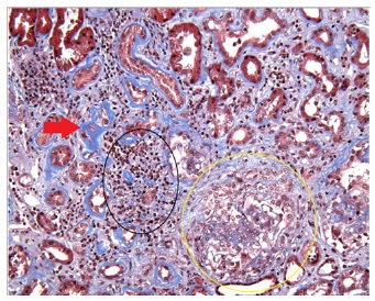
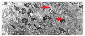
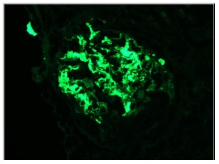
After correcting the patient’s electrolyte abnormalities, hemodialysis was initiated. Complement panel, antinuclear antibody, and antineutrophil cyctoplasmic antibody were all found to be noncontributory. Ultrasound imaging demonstrated bilateral echogenic kidneys of normal size. Despite undergoing repeated sessions of hemodialysis, the patient’s azotemia did not improve. On day 10 of admission, the patient underwent a renal core biopsy [10]; sections from one renal cortex containing 30 glomeruli were obtained. The sections demonstrated diffuse mesangial and focal segmental sclerosing glomerulonephritis, both acute and chronic. Thirteen glomeruli were globally sclerotic or approached global sclerosis. A portion of the sclerosing glomeruli demonstrated circumferential subcapsular fibrous proliferations with disruption of Bowman’s capsule, consistent with old fibrous crescents. Five glomeruli contained segmental scars with overlying segmental fibrocellular or fibrous crescents. The remaining glomeruli contained variable mild to moderate mesangial hypercellularity and increased matrix containing glassy mesangial eosinophilic and fuchsinophilic deposits. Seven glomeruli demonstrated segmental endocapillary proliferation, including focal infiltrating monocytes and neutrophils and irregular membranoproliferative features in the form of mesangial interposition and duplication of glomerular basement membranes superimposed on the mesangial proliferative features. Four glomeruli contained cellular crescents, three of which were segmental and one circumferential. The biopsy was also remarkable for severe tubular atrophy and interstitial fibrosis (60%) (Figure 1), diffuse, severe interstitial inflammation (80%) (Figure 1), and mild-to-moderate arterio- and arteriolosclerosis and hyalinosis. Though immunofluorescent sampling of eight glomeruli is typically preferred to establish diagnosis, the one glomerulus sampled in this instance demonstrated intense 3+ dominant granular global mesangial and segmental glomerular capillary wall positivity for IgA (Figures 3 and 4) with negative IgG along with other findings strongly supportive of the diagnosis of IgAN. These results along with the appearance of segmental crescents (Figure 2) indicate a poor prognosis as suggested by the Oxford classification of IgAN [11]: the glomerulus demonstrated diffuse mesangial proliferation (M1), focal endocapillary proliferation (E1), segmental sclerosis (S1), and >50% tubular atrophy/interstitial fibrosis (T2). Following the diagnosis, the patient was started on steroids and cyclophosphamide. Two months after discharge, mid-week BUN average was found to be 61mg/dl and creatinine was 6.5mg/dl. The patient is continuing on hemodialysis along with steroids and cyclophosphamide.
Figure 1: Trichrome stain demonstrating leukocyte infiltration (black oval) and mesangial
Figure 2: Reticulin stain demonstrating membranous deposits (red arrows) and a segmental cresent (yellow oval)
Figure 3: Electron microscopy demonstrating electron-dense mesangial deposits (red arrows)
Figure 4: Immunofluorescent glomerular stain demonstrating IgA antibody positivity
Discussion
Though a fairly common renal disorder globally, prevalence of IgAN among African Americans, particularly African American men, is comparatively low. It is worth noting, however, that a 1998 investigation of IgAN prevalance in central and eastern Kentucky demonstrated similar rates of IgAN in Caucasians and African Americans [12].
The constellation of atypical elements in this case, including the patient’s race and presentation—not gross hematuria but the less common presentation of microscopic hematuria, acute kidney injury, and nephrotic-range proteinuria— suggests that IgAN presentation among African Americans may be commonly atypical. There is abundant evidence suggesting that race can affect initial presentation and treatment response in kidney disease, particularly nephrotic syndrome. In addition to the well-documented racial disparities in IgAN prevalence, there is also evidence to suggest racial disparities in the response to treatment of IgAN. A 2006 study compared outcomes of Black and Indian children being managed with cyclophosphamide for steroid-resistant nephrotic syndrome and found substantial differences in the rates of remission between the two groups [13].
Future directions for research might include a race-based comparison of presenting complaints and urinalyses among confirmed cases of IgAN, as well as genetic and proteomic comparisons of African Americans and Caucasians that might shed light on hereditary factors protective against IgAN.
References
- D'Amico G (1987) The Commonest Glomerulonephritis in the World: IgA Nephropathy. Q J Med 64: 709-727.
- Galla JH (1995) IgA Nephropathy. Kidney Int 47: 377-387.
- Tumlin JA, Madaio MP, Hennigar R (2007) Idiopathic IgA Nephropathy: Pathogenesis, Histopathology, and Therapeutic Options. Clin J Am Soc Nephrol2: 1054-1061.
- Waldherr R (1989) Frequency of Mesangial IgA Deposits in a Non-selected Autopsy Series. Nephrol Dial Transplant 4: 943-946.
- Topham PS (1994) Glomerular Disease as a Cause of Isolated Microscopic Haematuria. Q J Med87: 329-335.
- Donadio JV (2002) IgA Nephropathy.N Engl J Med 347: 738-748.
- Mustonen J (1983) The Nephrotic Syndrome in IgA Glomerulonephritis: Response to Corticosteroid Therapy." Clin Nephrol 20: 172-176.
- Ibels LS, Györy AZ (1994) IgA Nephropathy: Analysis of the Natural History, Important Factors in the Progression of Renal Disease, and a Review of the Literature." Medicine (Baltimore) 73: 79-102.
- Jennette JC, Wall SD, Wilkman AS (1985) Low Incidence of IgA Nephropathy in Blacks. Kidney Int 28: 944-950.
- Neelakantappa K, Gallo GR, Baldwin DS (1988) Proteinuria in IgA Nephropathy. Kidney Int33: 716-721.
- Working Group of the International IgA Nephropathy Network and the Renal Pathology Society, Cattran DC, Coppo R, Cook HT, Feehally J, et al. (2009) The Oxford Classification of IgA Nephropathy: Rationale, Clinicopathological Correlations, and Classification. Kidney Int 76: 534-545.
- Wyatt RJ, Julian BA, Baehler RW, Stafford CC, McMorrow RG, et al. (1998) Epidemiology of IgA Nephropathy in Central and Eastern Kentucky for the Period 1975 through 1994. Central Kentucky Region of the Southeastern United States IgA Nephropathy DATABANK Project. J Am Soc Nephrol 9: 853-858.
- Bhimma R, Adhikari M, Asharam K (2006) Steroid-resistant Nephrotic Syndrome: The Influence of Race on Cyclophosphamide Sensitivity. Pediatr Nephrol 21: 1847-1853.


